Shoulder Surgery

Fixation of Proximal Humerus (Shoulder) Fractures

Fixation of Proximal Humerus (Shoulder) Fractures

Proximal humerus fractures are very common and in most cases can be managed without surgery. When surgery is indicated, it requires careful planning using 3D CT scans, meticulous surgical fracture fixation and close follow up with guided rehabilitation. Patients stay in hospital overnight and wear a sling for 6 weeks; strengthening is commenced at 3 months postoperatively. Surgery is able to produce a more reliable outcome for younger patients and those with higher demands in function and sport.

The decision to proceed with surgery is unique to each individual and based on a number of factors, such as fracture pattern, occupation, sporting demands, social situation and overall expectations.

The goal of proximal humerus (shoulder) fracture surgery are to obtain anatomical reduction and stable fixation to allow early range of motion and return to full function in a timely manner.

There are many parts to a proximal humerus (shoulder) fracture that need to be accounted for – indeed proximal humerus fracture surgery is complex and quite intricate. The different factors to consider are:

Humeral head (ball) fracture

  • The fracture usually occurs well below the head (surgical neck)
  • In some cases (<1%) it occurs higher in the head (anatomical neck). These fractures present a unique challenge as the “bone death” rate is higher

Humeral shaft (arm bone) fracture

  • There may be fracture extension down the shaft (arm)
  • If this is the case a longer plate may be needed. In some cases, a “rod” is preferred, which is placed inside the bone canal

Tuberosities (rotator cuff insertion points)

  • This is a critical part of the fracture surgery
  • The rotator cuff tendons attach circumferentially around the humeral head and are a constant deforming force (the muscles and tendons are pulling the fracture fragments away)
  • These forces need to neutralised with sutures which are repaired to the plate
  • Reattaching the tuberosities (with the rotator cuff) allows your shoulder to function

Biceps tendon

  • This tendon runs in the front of the shoulder and is almost always a source of pain after this fracture – so we routinely move this tendon to a better place to reduce your shoulder pain post-surgery.

Bone loss

  • In higher energy cases, there may be bone loss under the humeral head (ball); in these cases, Dr Pant may suggest taking bone from your pelvis and using it as a “strut” under the humeral head to improve your chance of fracture healing.

Blood supply

  • During your injury, shoulder reduction manoeuvre, and surgical exposure/fixation there is a risk the blood vessels that supply the humeral head may be injured or damaged.
  • The procedure is under a General Anaesthetic where you will be positioned on your back
  • The shoulder is then prepared with a special antiseptic solution (dark pink in colour) and the surgical drapes are applied to maintain a sterile field at all times.
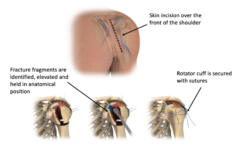
  • A 10 cm incision is made in the front of the shoulder and the planes are developed to find front of the shoulder joint; the fracture is then carefully defined and the fragments of bone are mobilised and the rotator cuff tendons are tagged with sutures (8-10 sutures).
  • The Biceps tendon at the front of the shoulder is then taken out of the shoulder joint and later re-anchored to the front of the humerus bone
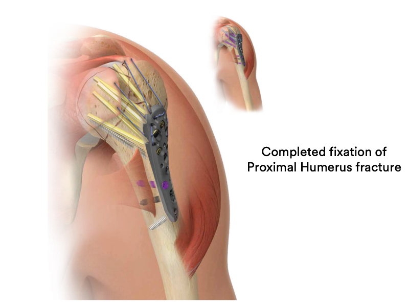
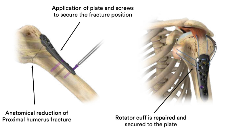
  • The fracture is then reduced and held in position with plates and screws
  • The rotator cuff tendons are sutured/repaired to the plate
    During the procedure, X-rays are taken to visualise the correct reduction and fixation
  • The wound is then closed in layers and dressings are applied; the arm is placed in a sling – either a standard sling or an abduction sling.
  • Dr Pant will then organise close follow up at regular intervals and liaise with your Physiotherapist to maximise your recovery
  • In <5% of cases Dr Pant may need to take bone from your pelvis to support the fracture position in your shoulder – if this might be the case for you, he will discuss this prior to surgery.

A series of 4 medical images showing the various stages of a open reduction and internal fixation surgery to repair a proximal humerus fracture

A series of 2 images showing the final stages of proximal humerus fracture repair surgery

A medical diagram showing a completed proximal humerus fracture repair surgery

Most patients report a sense of “relief” after their proximal humerus fracture is fixed and can commence light activity with the operated arm (such as self care, feeding, and using a mobile phone) immediately.

There are three essential components to a good outcome from surgery:

  • Your surgeon
  • You as a patient
  • Your physiotherapist

Post-operative physiotherapy

Dr Pant utilises the JPL pathway for most patients and this will form the basis of your rehabilitation after surgery; it will be modified to suit your individual circumstance.

The JPL pathway allows for self-directed, early passive shoulder range of movement:

  • Passive = assisted with your other arm
  • Active = you move the affected arm independently

Either a “standard” or an “abduction” sling is required for the first 6 weeks after your surgery. A “standard” sling is worn for a further 6 weeks outside of the house (to remind others you have had shoulder surgery). The type of sling is selected specific to you and your shoulder surgery.

Therapy overview 

After the surgery you will be in a sling and protect your shoulder to allow the shoulder fracture repair to heal. At two weeks after the surgery Dr Pant will see you at your review appointment and discuss the JPL rehabilitation protocol with you. This JPL physiotherapy protocol will commence two weeks after your surgery.

The therapy program will focus on flexibility and range of motion exercises initially. These gentle stretches will improve your range of motion and prevent shoulder stiffness. As the fracture begins to heal you will be able to progress to exercises that strengthen your shoulder muscles. Shoulder and rotator cuff strengthening exercises are commenced at 12 weeks after shoulder fracture surgery. Dr Pant will discuss when it is safe to return to your chosen sports. Generally, collision sports and overhead contact athletes need more time to allow for healing.

Driving after proximal humerus fracture surgery

  • After shoulder surgery, you will be in a sling and you may be unable to drive for at least 6-8 weeks

Showering and getting dressed

  • You may take the sling off to shower
  • Lean forward and allow your arm to “dangle” to wash under your arms
  • The dressings applied are waterproof and you may shower with them on
  • Usually, before you are discharged from Hospital, your dressings are changed after your morning shower.
  • These “new” dressings are then kept intact until your review at 10-14 days
  • If the dressings start to peel at the edges – you may reinforce them
  • If the dressings discolour with discharge (yellow or green) you may have a wound infection – Do not be concerned just yet; please notify SSU (02 9215 6100 or admin@drpant.com.au) and we will give you a plan.
  • When getting dressed, you may use your good arm to move your affected (operated arm) to place your arms through the sleeves carefully

Sleeping after Shoulder Surgery

  • Most patients find it difficult to lie flat after shoulder surgery (especially after an ORIF Proximal Humerus)
  • Consider using a few pillows to prop yourself up and sleep at a slight incline; this may be necessary for 4-6 weeks after surgery. You should keep your sling on while asleep.

Elbow and hand movement

  • Keep your elbow and hands joints moving and supple for the duration of time you are in the sling
  • The best time to move your elbow (into full extension and flexion) is when you are in the shower, just out of the shower and about to get dressed.
General Shoulder Surgery Risks

Surgery is a carefully choreographed process and you are being treated by a sub-specialist shoulder surgeon and a highly experienced team; however, all surgeries inherently carry some risk of complications.

The risk of complications after clavicle fracture surgery is less than 1% in the Sydney Shoulder Unit experience. General risks include:

  • Infection
  • Bleeding
  • Wound healing problems
  • Damage to blood vessels or nerves
  • Sensory changes around the surgical scar

Specific risks relating to proximal humerus fracture surgery

  • Delay in bone healing (delayed union)
  • Hardware complications (such as screw perforation of shoulder joint)
  • Neurovascular injury during surgery
  • Frozen shoulder
  • Avascular necrosis of humeral head (bone death and joint collapse)
  • Rotator cuff tear

Patients who smoke, use tobacco products, have diabetes, or are elderly are at higher risk of complications both during and after surgery. They are also more likely to have problems with wound and bone/tendon healing. Most healthy patients, however, cope well with shoulder fracture surgery and are unlikely to have complications.

The outcome after proximal humerus fracture surgery is good to excellent; however, it is not possible to restore the shoulder back to “normal”. During shoulder fracture surgery, your own damaged tissues are repaired in an attempt to restore your anatomy; often we can get this very close to normal/anatomical but the shoulder will unlikely return to its pre-injury state.

Most patients report a feeling of the shoulder being secure/stable immediately after surgery, and by two weeks they are back to basic things around the house. Therapy using the JPL protocol commences at two weeks post-surgery. At the final follow up visit at 3 months the fracture has usually healed and strengthening can commence. The chance of hardware complication in the Sydney Shoulder Unit experience is less than 5%.

Dr Pant has extensive experience treating proximal humerus fractures and is highly skilled in the surgical repair of all shoulder fractures. As the lead shoulder trauma surgeon at St Vincent’s Hospital for many years, he has experience with a vast array of shoulder fractures and is well versed in guiding you to full recovery.

Patient results

Shoulder Fracture – Proximal Humerus

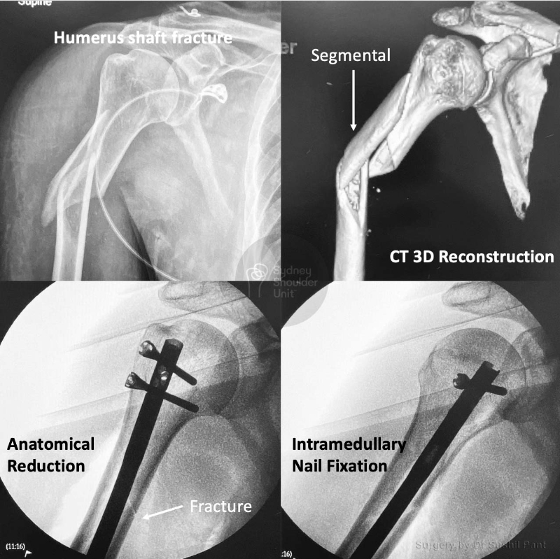
This is the case of a 50 year old man who sustained a torsional (twisting) injury to his right arm. He presented to hospital with pain and deformity to his right arm. Plain x-rays demonstrate a 3 part shaft fracture with comminution (multiple fragments). The CT scan 3D reconstructions show the extent of the fracture. The treatment advice was to proceed with surgery.

On this occasion, the fracture was reduced and fixed with a intramedullary nail (inside the bone) with locking screws on either end to stabilise the construct. The fracture line is now barely visible and he went on to unite his fracture and return to his previous level of function.

Shoulder Fracture – Gardening

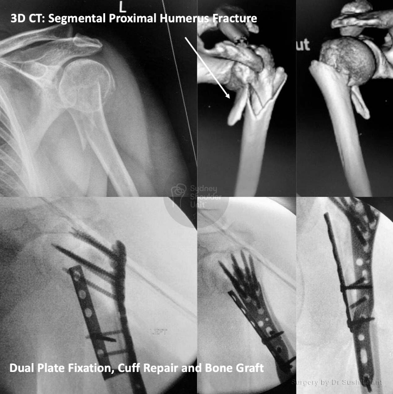
This 54 year old lady fell in her garden at home. She presented to the hospital with shoulder pain and swelling. She underwent immediate imaging with an x-ray and a CT scan with 3D reconstructions.

The fracture of the left shoulder was complex and shaft extension. This type of fracture is very difficult to fix as there is no medial hinge (due to the fractured shaft on the inside of the arm). She underwent an ORIF (Open Reduction and Internal Fixation) + bone graft + rotator cuff repair. The surgery was complex and needed a few tricks to get things looking perfect. Eventually, we achieved anatomical reduction and stable fixation to allow her early range of motion.

She commenced JPL rehabilitation at 2 weeks and united her fracture at 4 months post-surgery. She is back to gardening with new-found enthusiasm.

Shoulder Fracture – Scooter accident

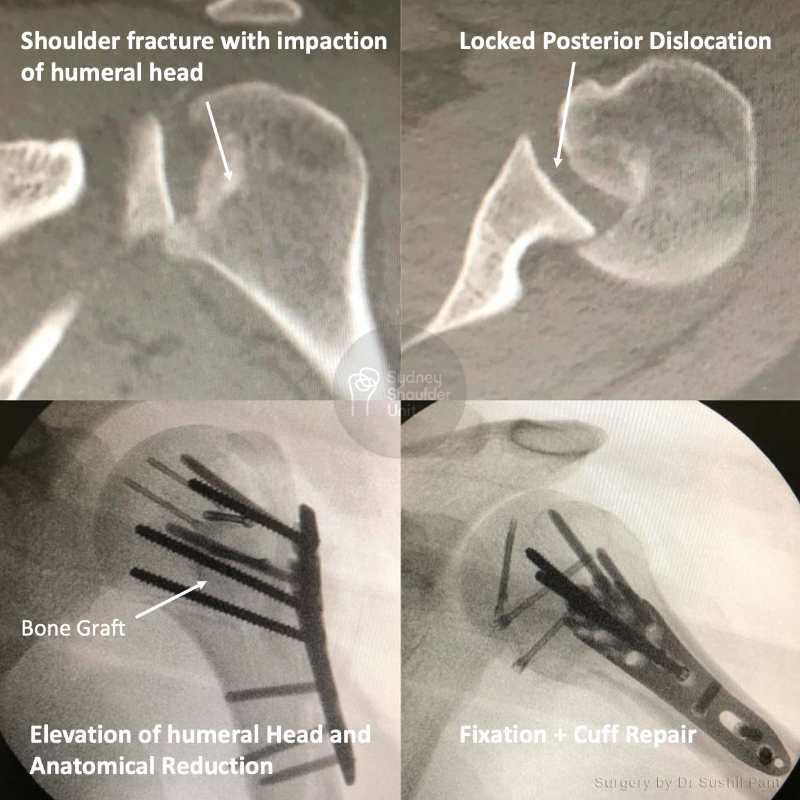
This 28 year old student had a scooter accident, sustaining this complex injury: a fracture with posterior dislocation and humeral head impaction resulting in articular cartilage injury.

He underwent an ORIF with humeral head elevation, iliac crest (pelvic bone) bone grafting and rotator cuff repair. The x-rays show the anatomical restoration of the humeral head articular surface with stable fixation. He was delighted with his eventual outcome given the complexity of his injury.

Shoulder Fracture – Displaced Humerus Fracture

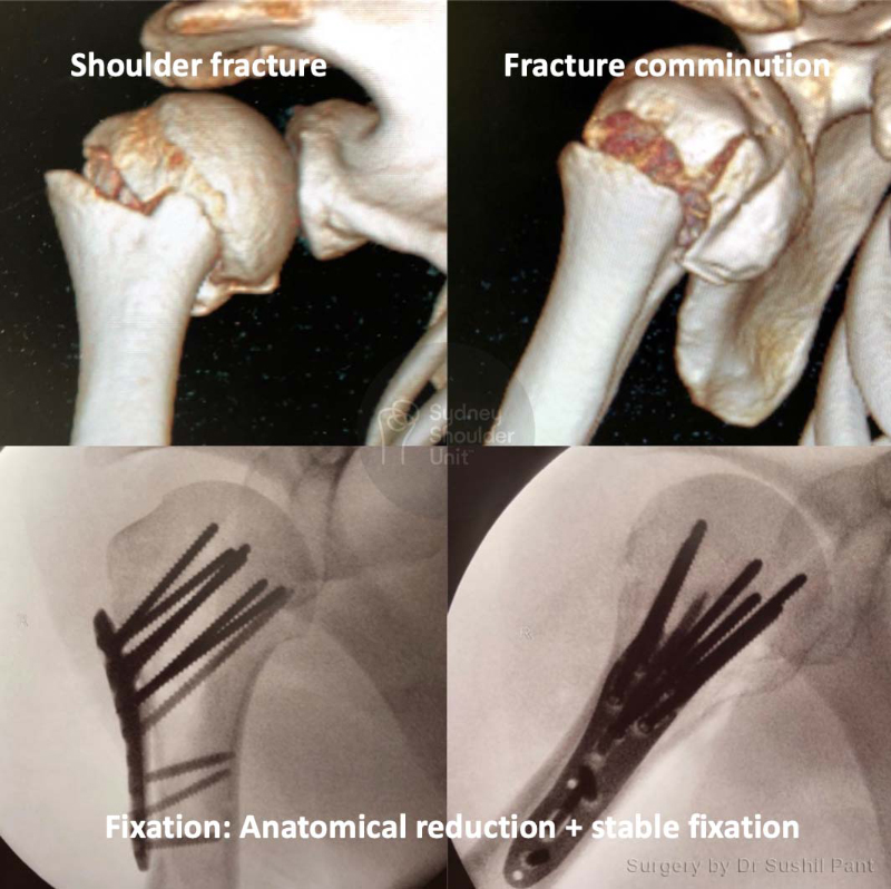
This 45 year old gentleman fell off a ladder at home sustaining this injury. The 3D reconstructions of the CT scan show a displaced proximal humerus (shoulder joint) fracture. This was fixed carefully using plates and screws to achieve anatomical realignment and stable fixation. He underwent JPL rehabilitation and then physiotherapy for strengthening, regaining normal, symmetrical range of motion and full function.

Shoulder Fracture – Complex Humerus Fracture

This lovely 63 year old lady fell at home, sustaining a very complex head split type of proximal humerus fracture. This case was borderline unreconstructable and an immediate shoulder replacement may be another option. After discussion with the patient, we elected to repair the shoulder fracture to allow her the best chance at keeping her own shoulder joint.

She underwent an ORIF (Open Reduction and Internal Fixation) and cuff repair. The pictures demonstrate the complexity of her fracture pattern. We achieved anatomical reduction and stable fixation: the fracture lines are barely visible on her post-surgical scans. After a period of therapy she went on to do very well regaining close to full function.

Request a call back

Enquiries between 8am-6pm (Mon-Fri) will be responded to within 30-60 minutes.

"*" indicates required fields

Alternatively, if you have any further questions or would like a consultation with Dr Pant get in touch:

Medicare Card Help

1. Medicare Number
2. Position on card
3. Expiry Date

Request a call back