Injuries and conditions

Biceps Tendonitis

Biceps Tendonitis

Biceps tendonitis often occurs in patients between the ages of 30-50 and typically presents with pain over the front of the shoulder. Patients usually report difficulty with activities overhead, reaching behind their back and clicking/catching at the front of the shoulder.

Biceps tendonitis often occurs with other conditions in the shoulder and therefore it requires careful workup and investigation to exclude SLAP tears, supraspinatus and subscapularis tendon tears, subacromial bursitis, and AC joint arthritis.

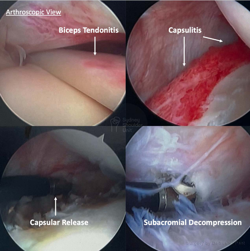
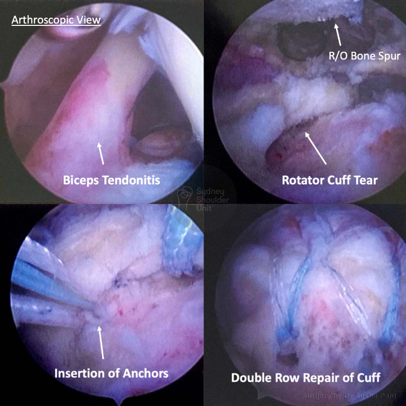
Biceps tendonitis is inflammation or irritation of the upper biceps tendon near the front of your shoulder. Also referred to as the “long head of the biceps tendon” (LHBT), this tendon is a strong cord-like structure that connects the biceps muscle to the top of the shoulder socket.

A common presentation of biceps tendonitis is pain at the front of the shoulder and weakness with certain shoulder movements. This is because the biceps tendon runs in a very narrow groove at the front and with inflammation may thicken and become irritated or even compressed. The biceps tendon normally glides about 2 cm during overhead shoulder movement.

In the early stages, the tendon becomes inflamed and swollen. With ongoing tendonitis, the tendon sheath (covering) can thicken, and eventually, the tendon itself may enlarge and become double or triple the size of a normal tendon. This enlarged tendon can be a source of pain over the front of the shoulder.

In some cases, the biceps tendon may tear (partial or complete). A complete tear will result in tendon rupture and loss of contour about the front of the arm, this is referred to as a “Popeye” sign.

Medical diagram showing the anatomy of the shoulder joint pointing out a thickened and enlarged Biceps tendon

In most cases biceps tendonitis is due to normal “wear and tear” over a normal lifetime of use. As we age, with repeated use, our tendons slowly weaken and degenerate; this may be worsened by overuse, particularly repeating the same shoulder motion again and again.

Overhead manual workers and sports persons are at particular risk. Sports, especially those that require repetitive overhead motion, such as swimming, tennis, and contact sport can put people at risk for biceps tendonitis.

Repetitive overhead motion may also play a part in other shoulder problems that may occur with biceps tendonitis or in isolation such as, shoulder impingement syndrome, subacromial bursitis, rotator cuff tears, osteoarthritis of the shoulder, and chronic shoulder instability.

During a comprehensive shoulder examination there are particular areas of pain and special tests that isolate the biceps tendon and help with the diagnosis.

  • Pain or tenderness at the front of the shoulder, which is aggravated with overhead motion and lifting
  • Pain or an ache that moves down the arm
  • Occasional snapping sound or sensation in the shoulder
  • Difficulty with putting on a seat belt, tucking a shirt in, or putting on a bra are some examples of tasks that are difficult with biceps tendonitis.

There are often other areas of pain generation within the shoulder that coexist and these should be identified and treated if present.

Imaging tests that are useful in the diagnosis of biceps tendonitis include a plain X-ray, Ultrasound and MRI scans. The MRI is particularly useful in looking for any evidence of biceps tendon tears, inflammation, biceps sheath fluid, and any other associated conditions such as rotator cuff tears and subacromial bursitis.

The majority of biceps tendonitis can be managed nonsurgically with rest, activity modification, and non-steroidal anti-inflammatory drugs (NSAIDs), and physiotherapy. An ultrasound guided cortisone injection is very effective with both diagnosis and treatment of biceps tendonitis. Such injections may provide relief for anwyere between a few weeks to a few months. Generally it is best not to have more than two injections per year as the cortisone may weaken the biceps tendon and lead to tendon rupture.

If your condition does not improve with nonsurgical treatment, then surgery may be an option for you. Surgery may also be a better option if you have other associated shoulder problems, particularly those that are time urgent.

Surgery for biceps tendonitis is performed arthroscopically (keyhole); this allows your surgeon to assess the condition of the biceps tendon and other structures in the shoulder.

Options at surgery:

  • Biceps tendon repair (SLAP repair): Typically suited to younger patients where the tendon itself is found to be in good condition at surgery but torn off the top of the glenoid (socket)
  • Biceps tenodesis: The damaged section of the tendon is removed and the remaining healthy biceps tendon is reattached to the upper arm bone (humerus) with an incision under the armpit. This is a very successful operation and removing the damaged/inflamed biceps tendon usually resolves symptoms and restores normal function
  • Biceps tenotomy: In some cases it may be best to release the damaged biceps tendon from the shoulder joint without reattaching it, this is called a “biceps tenotomy”. This is ideal in the older patient and does not weaken shoulder or arm function; however, in some cases, it may leave a “Popeye” bulge over the arm. Patients who have a tenotomy for the right reasons do very well and return to normal shoulder function.

The key point is to remember which conditions in your shoulder are “time urgent” and which are not. Dr Pant will clearly outline this for you during your consultation.

Time urgent shoulder conditions

Biceps tendon dislocation (out of the groove).

When the biceps tendon is “subluxed” or “dislocated” out of the groove it can cut into the subscapularis tendon and cause further damage. You should consider surgery sooner rather than later.

Non urgent considerations

Shoudler function

If you are unable to undertake basic tasks or to look after yourself, unable to drive, unable to get dressed, or have difficulty with hygiene and toileting.

Shoudler pain

  • Pain with activity and even at rest
  • Night pain, especially if it wakes you from sleep
  • Unable to sleep on your affected shoulder
  • Increasing requirements of pain relief tablets
  • Multiple failed cortisone injections
  • Not responding to physiotherapy

Work

  • If it is limiting your ability to work
  • Especially if you are in a manual job requiring repeated overhead activity

Sport

If you are unable (even after a period of rest and activity modification)  to participate in your chosen leisure activity or sport and you wish to continue to do so.

Prevent further shoulder damage

There are certain conditions where it may be better to repair your shoulder before further damage occurs. Dr Pant will carefully analyse your MRI scan and determine if this is the case for you (especially in younger patients).

Patient results

Frozen Shoulder Release

This gentleman in his 60s suffered from a frozen shoulder for almost a year; his symptoms were unrelenting and he failed to improve over time despite non-surgical treatment. Examination and MRI imaging confirmed ongoing frozen shoulder as well as biceps tendonitis, subacromial bursitis, and AC joint arthritis; thankfully his rotator cuff was intact. 

He underwent arthroscopic frozen shoulder surgery (capsular release) and a biceps tenodesis, subacromial decompression and excision of his AC joint. Range of motion exercises were commenced immediately after surgery using the JPL protocol.

Rotator Cuff Tear

This 53 year old lady presented with right-sided shoulder pain after injuring her shoulder at work. She underwent a period of rest, cortisone injection and 3 months of physiotherapy without improvement.

After a careful history and examination, an MRI scan was undertaken. The MRI showed a Rotator Cuff Tear with Biceps Tendonitis.

The images taken at surgery (key-hole) demonstrate significant biceps tendonitis with inflammation and thickening of the tendon. The biceps tendon was taken out of the shoulder joint and re-anchored under the arm-pit (biceps tenodesis). The bursitis and bone spur was cleaned out and the rotator cuff tear was repaired with 4 anchors with good compression of the tendon foot-print.

She was discharged home the following day after surgery and placed in a sling for six weeks. She underwent JPL rehab which commenced at two weeks post-surgery. Strengthening commenced at months under the guidance of her physiotherapist.

She no longer has her previous shoulder pain and has good power of her rotator cuff.

Request a call back

Enquiries between 8am-6pm (Mon-Fri) will be responded to within 30-60 minutes.

"*" indicates required fields

Alternatively, if you have any further questions or would like a consultation with Dr Pant get in touch:

Medicare Card Help

1. Medicare Number
2. Position on card
3. Expiry Date

Request a call back