Injuries and conditions

Proximal Humerus (Shoulder) Fractures

Proximal Humerus fractures

Fractures around the shoulder may occur due to a simple fall, playing sport, or in a motor vehicle accident. These fractures are very common and need careful assessment with X-ray, CT and/or MRI scans. It is important to understand the exact pattern of the fracture and the patients expectations around fracture healing and recovery. Any other associated injuries should be identified, such as rotator cuff tendon tears. Most patients can be managed without surgery; if surgery is required it is best to undertake that sooner rather than later.

The proximal humerus refers to the upper arm bone as it becomes the ball of the shoulder joint. Hence proximal humerus fractures are fractures that involve the shoulder joint.

There are different parts of the ball that may fracture, sometimes it is just one of these parts, other times it’s all four parts. With significant force the shoulder may dislocate out of the socket (this is known as a fracture-dislocation).

  • Humeral head (ball of the shoulder joint)
  • Greater tuberosity (the outside part of the ball where the supraspinatus and infraspinatus tendon attach)
  • Lesser tuberosity (the inside part of the ball where the subscapularis tendon attaches)
  • Shaft (the top of the arm bone, humerus)

Medical diagram showing the anatomy of the shoulder joint with a Proximal Humerus Fracture

Proximal humerus fractures occur in two types of patients: those who are older with osteoporotic bone (low energy trauma) and younger patients with good quality bone involved in high energy trauma.

Fractures of the proximal humerus may occur due to a simple fall, during sport, at work, or because of a higher energy mechanism (such as from a bicycle or motor vehicle).

Most patients present to the hospital with pain and loss of function around the shoulder and arm bone after an injury. The diagnosis is suspected clinically and confirmed on plain X-ray. In many cases, a CT scan is needed to assess the extent of the fracture, the position of the fragments and other associated injuries. It is important to carefully examine the arm for any neurological or vascular issues.

Xray and 3D CT Scan showing a shoulder joint with Proximal Humerus Fracture

Most fractures of the shoulder (proximal humerus or surgical neck of humerus) can be managed without surgery using a simple sling. These patients are followed with imaging and the position of the fracture is monitored as the bones heal over time (3 months). The younger the patient and the higher their sporting or work demands the more likely they are to benefit from surgical fixation.

Non-surgical treatment for proximal humerus fractures consists of:

  • Use of a sling for 6 weeks
  • Pain relief
  • Follow up at 1, 2, 6 and 12 weeks with routine X-rays to check for fracture displacement and/or evidence of fracture healing

Surgical fixation is reserved for more complex cases such as fractures with displacement (fracture fragments shifted out of position), high demand patients (overhead workers, high-level athletes), or those that are unable to cope with the pain or loss of function.

Dr Pant has extensive experience treating proximal humerus fractures and is highly skilled in the surgical repair of all shoulder fractures should that be required. As the lead shoulder trauma surgeon at St Vincent’s Hospital for many years, he has experience with a vast array of shoulder trauma cases and is well versed in guiding you to full recovery.

These are the factors when considering surgery:

1.  Fracture displacement

If the fracture fragments are too far apart then they may heal in the wrong position; once the fracture has healed it is often very difficult to “re-break” and “reset” them in the correct position. Abnormal fracture position may result in reduced shoulder function.

After the first 2-4 weeks, the fracture will start to heal and will become difficult to mobilise and fix accurately.

2. Tuberosity and Rotator cuff involvement

If the tuberosities (rotator cuff attachment points on the humeral head) have pulled away more than 5-10mm then you may benefit from surgery. These fragments have a constant deforming force and will usually continue to move further away (over days to weeks) while the fracture is healing.

3. High-level athletes

In these cases, surgery is a better option early to allow faster recovery and more reliable outcomes. Tuberosity displacement is accepted up to 3mm.

4. Overhead work

Those patients who depend on overhead function at work may benefit from earlier surgery to minimise the time in a sling, shoulder stiffness, and enhance their ability to undertake work overhead.

5. Other issues

Some patients may live alone, be a carer for their significant other, be unable to tolerate six weeks in a sling, or unable to manage their pain – in such cases it is appropriate to consider surgical repair to allow more predictable and faster recovery.

Patient results

Shoulder Fracture – Proximal Humerus

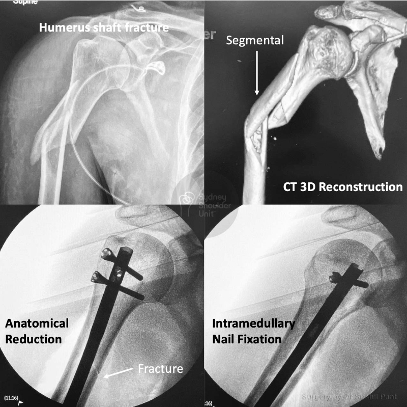
This is the case of a 50 year old man who sustained a torsional (twisting) injury to his right arm. He presented to hospital with pain and deformity to his right arm. Plain x-rays demonstrate a 3 part shaft fracture with comminution (multiple fragments). The CT scan 3D reconstructions show the extent of the fracture. The treatment advice was to proceed with surgery.

On this occasion, the fracture was reduced and fixed with a intramedullary nail (inside the bone) with locking screws on either end to stabilise the construct. The fracture line is now barely visible and he went on to unite his fracture and return to his previous level of function.

Shoulder Fracture – Gardening

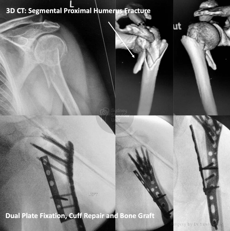
This 54 year old lady fell in her garden at home. She presented to the hospital with shoulder pain and swelling. She underwent immediate imaging with an x-ray and a CT scan with 3D reconstructions.

The fracture of the left shoulder was complex and shaft extension. This type of fracture is very difficult to fix as there is no medial hinge (due to the fractured shaft on the inside of the arm). She underwent an ORIF (Open Reduction and Internal Fixation) + bone graft + rotator cuff repair. The surgery was complex and needed a few tricks to get things looking perfect. Eventually, we achieved anatomical reduction and stable fixation to allow her early range of motion.

She commenced JPL rehabilitation at 2 weeks and united her fracture at 4 months post-surgery. She is back to gardening with new-found enthusiasm.

Shoulder Fracture – Scooter accident

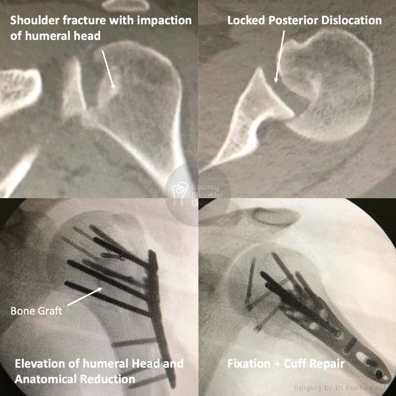
This 28 year old student had a scooter accident, sustaining this complex injury: a fracture with posterior dislocation and humeral head impaction resulting in articular cartilage injury.

He underwent an ORIF with humeral head elevation, iliac crest (pelvic bone) bone grafting and rotator cuff repair. The x-rays show the anatomical restoration of the humeral head articular surface with stable fixation. He was delighted with his eventual outcome given the complexity of his injury.

Shoulder Fracture – Displaced Humerus Fracture

This 45 year old gentleman fell off a ladder at home sustaining this injury. The 3D reconstructions of the CT scan show a displaced proximal humerus (shoulder joint) fracture. This was fixed carefully using plates and screws to achieve anatomical realignment and stable fixation. He underwent JPL rehabilitation and then physiotherapy for strengthening, regaining normal, symmetrical range of motion and full function.

Shoulder Fracture – Complex Humerus Fracture

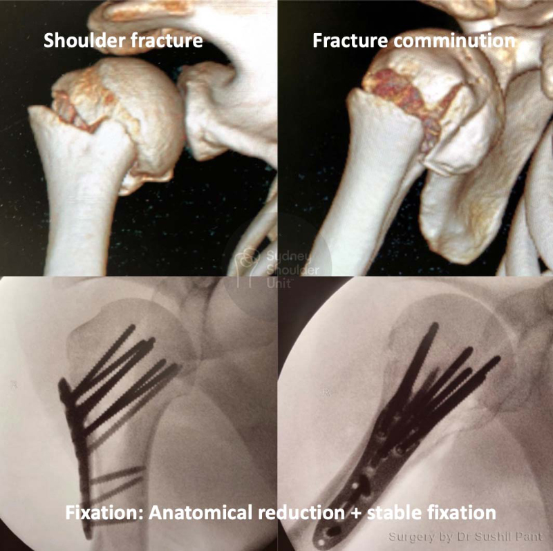
This lovely 63 year old lady fell at home, sustaining a very complex head split type of proximal humerus fracture. This case was borderline unreconstructable and an immediate shoulder replacement may be another option. After discussion with the patient, we elected to repair the shoulder fracture to allow her the best chance at keeping her own shoulder joint.

She underwent an ORIF (Open Reduction and Internal Fixation) and cuff repair. The pictures demonstrate the complexity of her fracture pattern. We achieved anatomical reduction and stable fixation: the fracture lines are barely visible on her post-surgical scans. After a period of therapy she went on to do very well regaining close to full function.

Request a call back

Enquiries between 8am-6pm (Mon-Fri) will be responded to within 30-60 minutes.

"*" indicates required fields

Alternatively, if you have any further questions or would like a consultation with Dr Pant get in touch:

Medicare Card Help

1. Medicare Number
2. Position on card
3. Expiry Date

Request a call back