Injuries and conditions

Calcific Tendonitis

Calcific Tendonitis

Calcific tendonitis most commonly affects people between the ages of 40- 60, and occurs in women more often than men (2:1). The cause of calcific tendonitis is not known and patients usually present with shoulder pain and stiffness. Initial treatment is usually non-surgical, with a period of rest, cortisone injection, anti-inflammatories, and guided physiotherapy. In about 10% of cases keyhole surgery may be required to remove the calcium deposit and expedite recovery, relieve pain and/or treat other coexisting conditions (such as a supraspinatus tendon tear).

Calcific tendonitis occurs when calcium deposition occurs near the rotator cuff insertion. This may be associated with bursitis and cuff degeneration. In some cases, there is no pain and this is an incidental finding on a plain shoulder X-ray. The calcium deposition most commonly occurs in the supraspinatus tendon.

There are three stages of calcific tendonitis: 

  1. Pre-calcific: pain with movement and reduced shoulder movement
  2. Calcific: cells release calcium which deposits in the tendon; after some time the body begins to resorb the calcium and this can cause significant pain and discomfort. 
  3. Post-calcific: the areas of calcium deposits are replaced with healthy tissue and range of motion beings to improve

Patients often present with shoulder pain and a reduced range of motion.

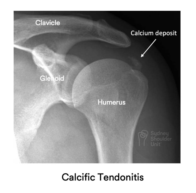
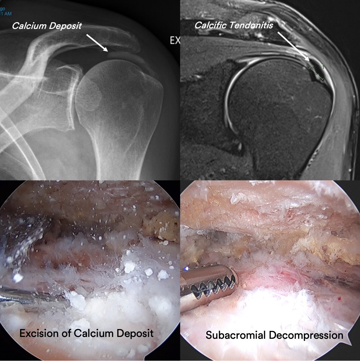
The diagnosis is often made with a plain x-ray where a calcium deposit may be seen above the tendon insertion. In some cases, it is prudent to obtain an MRI scan to exclude a rotator cuff tear or other pathologies about the shoulder. A supraspinatus tendon tear is not uncommonly associated with calcific tendonitis.

Xray image of shoulder joint showing Calcific Tendonitis

The initial days of calcific deposit formation in the tendons may be very painful and patients may require cortisone for relief. Most cases of calcific tendonitis are self-limiting and treatment is usually a course of NSAIDs, physiotherapy, corticosteroid injections into the bursa. In some cases ultrasound-guided needling and lavage is beneficial.

Surgery is required in about 10% of cases and involves an arthroscopic removal of the calcium deposit and subacromial bursectomy + acromioplasty. This is reserved for patients with recalcitrant symptoms or underlying rotator cuff tears (where the cuff tear may benefit from repair – such as a supraspinatus tendon tear).

Patient results

Calcific Tendonitis

This 59 year old gentleman presented with unrelenting shoulder pain which was progressively getting worse.  He reported no prior trauma and was finding it difficult to undertake activities of daily living and unable to jog. He required pain killers and elected to proceed with key-hole surgery given his lack of improvement with non-surgical treatment.

He underwent an Arthroscopy (Key-hole) of the left shoulder and the calcium deposit in the Supraspinatus Tendon was removed entirely. The tendon was stable at the end of surgery and did not require treatment. He used a sling for 2 weeks and commenced immediate range of motion as tolerated using the JPL protocol.


Request a call back

Enquiries between 8am-6pm (Mon-Fri) will be responded to within 30-60 minutes.

"*" indicates required fields

Alternatively, if you have any further questions or would like a consultation with Dr Pant get in touch:

Medicare Card Help

1. Medicare Number
2. Position on card
3. Expiry Date

Request a call back