
Arthroscopic excision of calcium is a minimally invasive keyhole procedure to treat calcific tendonitis. It is often done as a part of other keyhole procedures in the shoulder, such as a subacromial decompression, biceps tenodesis and/or rotator cuff repair. After an isolated arthroscopic excision of calcium a sling is worn for 1-2 weeks; and JPL therapy commences immediately after surgery. Strengthening is allowed at 4-6 weeks; and full unrestricted activity is possible at 6-12 weeks after surgery. This pathway is modified if other procedures are carried out at the same time.
This is a keyhole procedure that uses cameras and small instruments inside the shoulder to remove the calcium deposit in the rotator cuff tendon (usually the supraspinatus tendon). There may also be a supraspinatus tendon tear which can be treated if needed at the same time. In some cases the biceps tendon and or the AC joint may also need treatment.
Physiotherapy is commenced 2 weeks after surgery and the procedure can be done as a day stay or with an overnight admission.
Although most patients improve with cortisone injection into the bursa and ultrasound guided needling, symptoms may persist in some patients.
Surgery is suitable for those patients who are not responding to non-surgical treatments and find that:
If your symptoms have persisted for six months and your condition is deteriorating then surgery is a good option (particularly for the younger patient).
There are three essential components to a good outcome from surgery
Dr Pant utilises the JPL pathway for most patients and this will form the basis of your rehabilitation after surgery.
The JPL pathway allows for self-directed, early passive shoulder range of movement:
A sling may be worn for up to 6 weeks after surgery. However, after isolated arthroscopic excision of calcium surgery, Dr Pant will encourage you to come out of the sling as much as possible – to commence early physiotherapy immediately after surgery.
Surgery is a carefully choreographed process and you are being treated by a sub-specialist shoulder surgeon and a highly experienced team; however, all surgeries inherently carry some risk of complications.
The risk of complications after isolated arthroscopic excision of calcium surgery are less than 1% in the Sydney Shoulder Unit experience.
General risks:
Specific risks relating to Arthroscopic excision of calcium surgery:
Patients who smoke, use tobacco products, have diabetes, or elderly are at higher risk of complications both during and after surgery. They are also more likely to have problems with wound healing.
With careful patient selection, surgical removal of the calcium deposit and early physiotherapy patients report improved range of motion and less pain.

Dr Sushil Pant is a leading Australian trained orthopaedic shoulder surgeon. He is the founder and medical director of the Sydney Shoulder Unit; and is a Shoulder Surgeon at Sydney Sports Medicine Centre at Sydney Olympic Park.
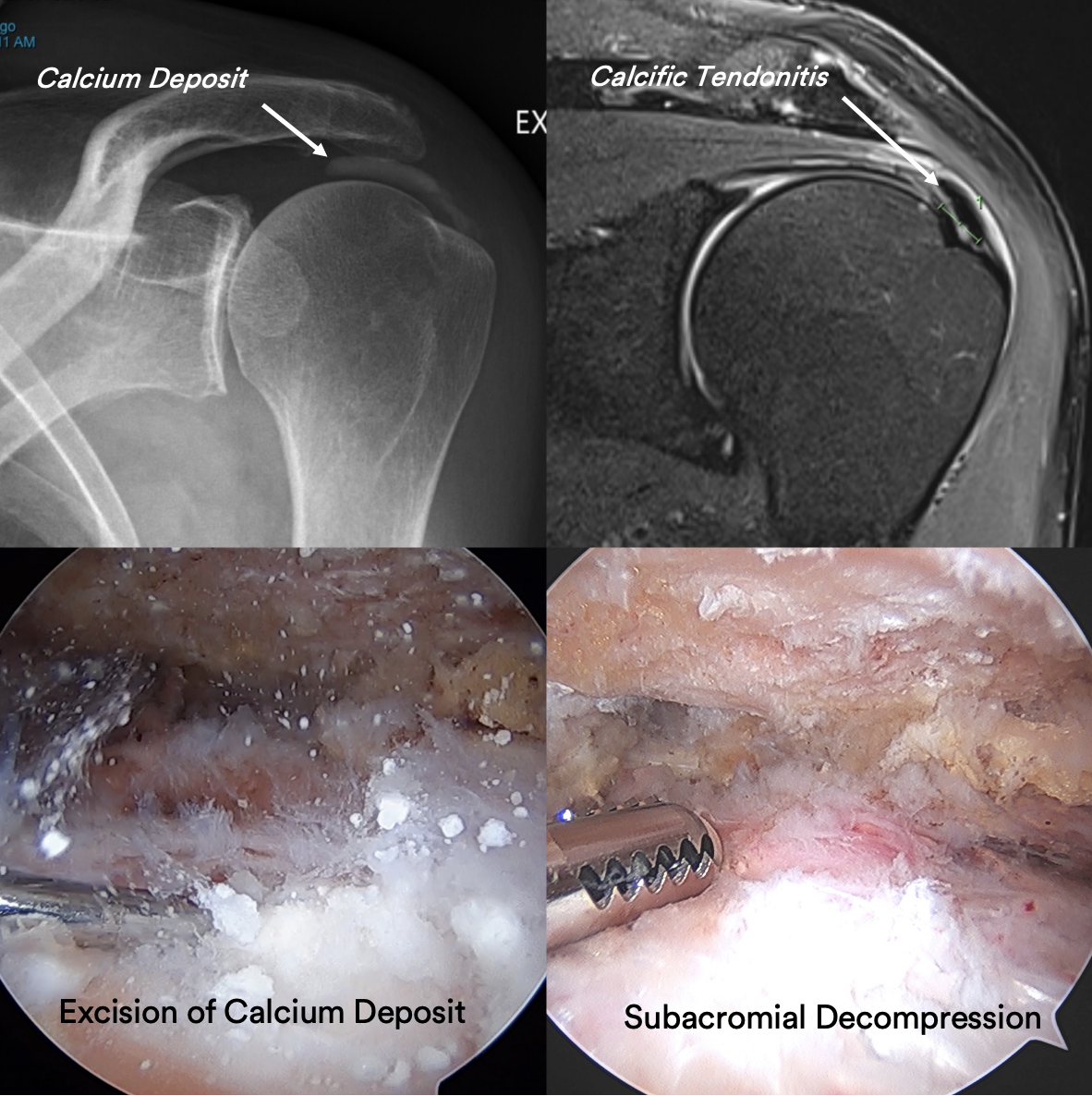
This 59 year old gentleman presented with unrelenting shoulder pain which was progressively getting worse. He reported no prior trauma and was finding it difficult to undertake activities of daily living and unable to jog. He required pain killers and elected to proceed with key-hole surgery given his lack of improvement with non-surgical treatment.
He underwent an Arthroscopy (Key-hole) of the left shoulder and the calcium deposit in the Supraspinatus Tendon was removed entirely. The tendon was stable at the end of surgery and did not require treatment. He used a sling for 2 weeks and commenced immediate range of motion as tolerated using the JPL protocol.
Enquiries between 8am-6pm (Mon-Fri) will be responded to within 30-60 minutes.
"*" indicates required fields
Alternatively, if you have any further questions or would like a consultation with Dr Pant get in touch:
1. Medicare Number
2. Position on card
3. Expiry Date