Injuries and conditions

Shoulder Joint Arthritis

Shoulder Joint Arthritis

Shoulder arthritis usually starts off as inflammation, over time it leads to loss of cartilage and joint space narrowing; this in turn leads to pain and stiffness which can make it difficult to lift your arm, brush your hair, reach up to a high shelf, and even self care and hygiene.

There is no cure for arthritis, however, there are many non-surgical and surgical treatment options available. The surgical treatments may be either keyhole or open (joint replacement). Recent advances in 3D printing and computer guided surgery has resulted in improved outcomes for patients.

There are two main joints that may be affected by arthritis. The first is the acromioclavicular (AC) joint – where the end of the collarbone (clavicle) meets the roof the shoulder (acromion). The other is where the head of the humerus (ball) fits into the glenoid (shoulder blade socket) – this is called the glenohumeral joint.

When we generally speak about shoulder arthritis – it usually refers to “glenohumeral” joint arthritis.

Arthritis refers to a condition where the cartilage covering the surface of a joint wears away, this causes bone-bone contact which leads to loss of movement and pain.

The shoulder joint is surrounded by the rotator cuff muscles (four in total) and the deltoid muscle which functions to move the shoulder joint.

As we age the quality of the cartilage and tendons may degenerate leading to pain, stiffness and weakness of the shoulder joint.

2 medical diagrams showing the normal shoulder anatomy and healthy rotator cuff tendons

There are five major types of arthritis typically affect the shoulder joint:

Osteoarthritis

This is most common form of arthritis in the shoulder
It is also called “wear and tear” arthritis and this condition that destroys smooth outer covering of bone (as it forms a joint) – this is the articular cartilage. As the articular cartilage wears away the protective space between bones narrows and the bones of the joint rub against each other, causing pain. This is referred to as “bone on bone” arthritis. Osteoarthritis typically affect those over the age of 55

Rheumatoid Arthritis

Rheumatoid arthritis is a systemic inflammatory condition that affects the bones, tendons and joints. This chronic condition may affect multiple joints in the body. Uniquely Rheumatoid arthritis affects the synovium, the lining that covers all joints in the body which lubricates the joint and makes it easier to move. The synovium becomes thickened and inflamed and this leads to pain and swelling of the joint. Over time the inflammation will start to attack the articular cartilage

Rheumatoid arthritis is an autoimmune condition – which means that the immune system attacks its own tissues.

More recently medication (DMARDs) used to treat Rheumatoid arthritis have become advanced and this has meant that the rate of joint involvement has dropped markedly.

Posttraumatic arthritis

This is a form of osteoarthritis that develops after injury, such as a shoulder fracture or dislocation. It usually takes many years to develop after the initial trauma. Patients who develop posttraumatic arthritis tend to be younger, usually 40-50 year olds.

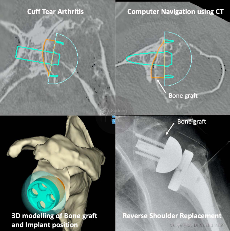
Rotator Cuff Tear Arthritis

Arthritis may also develop due to long-standing rotator cuff tendon tears. As the tendon tear becomes large, retracts and is irreparable the shoulder joint is no longer held in place and the ball moves up and out of the socket to sit under the acromion. This will lead to cartilage damage in the joint over time and is referred to as cuff tear arthropathy (arthritis).

The pain from arthritis (bone on bone) and loss of rotator cuff tendon function can render the arm very painful and without function – at the end stages it looks clinically like shoulder paralysis (referred to as pseudoparalysis).

Avascular Necrosis

Avascular necrosis (AVN) occurs when the blood supply to the humeral head is disrupted (that is it becomes avascular) and this causes bone cells to die (referred to as necrosis).

AVN progresses through stages – with progressive more bone death, cartilage damage, and joint collapse.

Initially, AVN starts off affecting the humeral head (ball) and over time it may also affect the glenoid (socket). The causes of AVN are:

  • High dose steroid use
  • Heavy alcohol consumption
  • Sickle cell disease
  • Trauma (such as fracture or dislocation)
  • Unknown (Idiopathic)

Series of 4 shoulder joint xrays showing Osteoarthritis, Cuff tear arthritis, Avascular Necrosis and Rheumatiod Arthritis

Diagnosis involves a combination of history, physical examination and imaging. The most common symptoms are:

Shoulder Pain

This is the most common symptom of arthritis; the pain is aggravated by activity and progressively worsens over time. It may also occur at rest and at night – waking patients up from sleep and making it difficult to sleep on the affected side.
The location of the pain may occur deep within the joint, over the front or back of the shoulder, over the deltoid, and sometimes down the arm or radiate up the neck.

Reduced range of motion

With progression of arthritis as the joint space narrows patients notice loss of range of motion. They may report limited external rotation and forward elevation – making it difficult to brush their hair, dress, self care, hang washing, and reach up to a shelf.

Crepitus

This occurs towards the end stages of arthritis – where grinding, clicking, snapping with shoulder movement is noted. This can cause pain and even be heard by others sometimes.

Examination

A thorough examination is undertaken to exclude other possible conditions and check the integrity of the rotator cuff tendons, deltoid muscle and nerves around the shoulder.

These include:

  • Inspection to look for previous surgery scars, skin changes and wasting of muscles
  • Palpation of the AC Joint, biceps tendon and shoulder joint lines
  • Movement (passive and active) of the shoulder is documented in all planes; any crepitus (joint nose) is noted
  • Power of the rotator cuff tendons and ligaments
  • Nerves of the shoulder joint
  • General health and any other joint involvement
    Imaging:

Plain X-ray

  • This is the first and easiest investigation to obtain; and in many cases can make the diagnosis.
  • It will also show a global view of the shoulder joint and exclude other conditions (such as bone cysts and tumours)
  • Arthritis of the shoulder will show narrowing of the joint; with advanced arthritis obliterating the joint completely – resulting in a bone on bone appearance.

Ultrasound

This is of limited value; however, it may provide some clues as to the integrity of the rotator cuff tendons.

CT scan

  • A CT scan will show the bony anatomy in 3D and will allow careful assessment of the joint wear pattern.
  • Sometimes it is required to confirm the diagnosis; but it is best reserved for pre-surgery planning. This is where the CT scan is uploaded to a special software and 3D measurements are taken of the patient’s shoulder, the exact wear pattern is analysed, and the correction is planned and the “virtual” prosthesis can be implanted on the computer. Then custom, patient-specific instruments are ordered for the day of surgery – the planned operation is then carried out with a high level of precision and accuracy.

MRI

In early cases of arthritis or AVN an MRI may be needed to make the diagnosis
However, this test is best for confirming the quality and integrity of the rotator cuff when a total shoulder replacement is being considered.

For the total shoulder replacement to function, it requires an intact rotator cuff. As opposed to a reverse shoulder replacement which can function without a rotator cuff (as the deltoid is the driving muscle force).

As with most arthritic conditions, the initial treatment is almost always non-surgical. The principles of non-surgical treatment are:

Rest and activity modification

You may need a short period of rest and/or change the way you use your arm to limit activities that exacerbate shoulder pain.

Analgesia

Non-steroidal anti-inflammatory (NSAIDs) and paracetamol are a good start to controlling baseline pain. Long-acting pain killers overnight can be useful. Prolonged use of NSAIDs may irritate the stomach lining and lead to gastric ulcers – therefore they should be used for short periods under the guidance of your local doctor.

Physiotherapy

Both self-directed and guided physical therapy is useful to keep the shoulder joint supple and strengthen the rotator cuff, scapula stabilising, upper back and deltoid muscles. This may offload the arthritis pain made worse by muscle imbalance.

Cortisone injections

Cortisone injections when used correctly can dramatically reduce inflammation and arthritis pain. However, this is not a cure and the effect is generally temporary. No more than two injections are recommended per year. A plain X-ray of the shoulder should always be obtained prior to cortisone injection into the joint – as cortisone itself is associated with avascular necrosis (AVN) of the shoulder joint – and this needs to be excluded prior to the injection.

General health optimisation 

Seeing your general practitioner and optimising your cardiovascular health, diabetes, blood pressure, and diet can all lead to improvement in overall health. It also makes you a better anaesthetic and surgical candidate should you require future surgery.

Surgical treatment

If your shoulder arthritis symptoms are not responding to non-surgical treatment then there are a number of surgical options:

Arthroscopy

In cases of mild shoulder arthritis, arthroscopic (keyhole) shoulder surgery may be used to target pain generators in the shoulder. These include:

  • Biceps tendon
  • Acromion spur and subacromial bursa
  • Acrmioclavicuar joint (ACJ)
  • Shoulder joint spurs

Arthroscopy will not eliminate arthritis, but treat areas that may be exacerbating your shoulder pain; this will delay the need for shoulder replacement surgery.
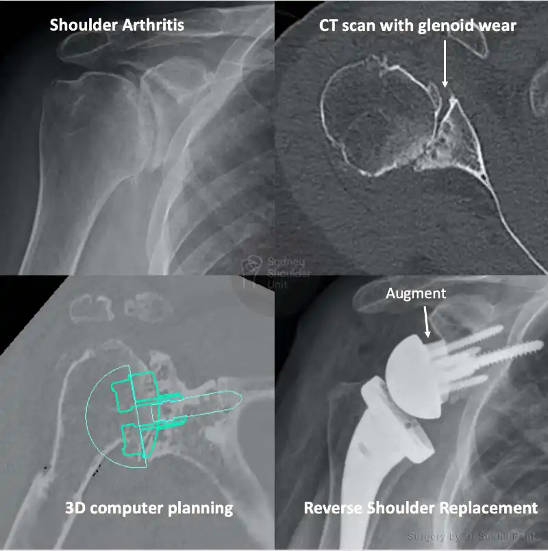
Joint Replacement

Partial joint replacement

  • Just the humeral head (ball) is replaced
  • This is uncommon and usually for younger patients and those with avascular necrosis (AVN)

Total joint replacement

Anatomical shoulder replacement

  • Both the head of the humerus and the glenoid (socket) are replaced
  • The head is replaced with a metallic ball
  • The glenoid is replaced with a plastic socket
  • The native anatomy and biomechanics of the shoulder are restored (hence this is called an anatomical shoulder replacement)
  • This does require an intact and good quality rotator cuff tendons (as assessed on MRI)

Reverse Shoulder Replacement

  • Both the head of the humerus and the glenoid (socket) are replaced
  • The head is replaced with a plastic socket
  • The glenoid is replaced with a metallic ball
  • The native anatomy and biomechanics of the shoulder are reversed (hence this is called a reverse shoulder replacement)
  • A reverse shoulder replacement does not require an intact rotator cuff; however, it does require a well functioning deltoid muscle.

2 medical diagrams comparing the difference between a total shoulder replacement and reverse shoulder replacement when treating shoulder joint arthritis

Many patients with shoulder arthritis can be managed without surgery. However, for those patients who have exhausted non-operative treatment, computer navigated 3D planning before shoulder surgery can produce reliable results and pain-free function.

Sydney Shoulder Unit is a high volume shoulder practice that performs a large number of shoulder replacements each year with consistent outcomes for our patients.

For patients who have no rotator cuff and advanced arthritis, the Reverse Shoulder Replacement has offered new treatment options.

The latest advances in computer navigation, 3D planning and printing, and patient-specific instrumentation allow for accurate placement of components resulting in superior radiographic and functional outcomes for our patients.

Australian Orthopaedic Association National Joint Replacement Registry Data (Individual Surgeon Reports) shows that Dr Pant’s revision rate for shoulder replacements is currently 0.00% (as of 2021); the national average is 1.06%. This includes Total Shoulder Replacements, Reverse Shoulder Replacements and Shoulder Replacements for fractur

Patient results

Total Shoulder Replacement

This 70 year old lady presented with many years of shoulder pain. With the pain affecting her sleep and ability to undertake activities of daily living. Having trailed multiple cortisone injections over the years and an intense program of physiotherapy, she was at the end of her tether. 

Plain x-rays demonstrated end stage advanced osteoarthritis. MRI scan demonstrated an intact Rotator cuff – which is required for the “anatomical” total shoulder replacement to function well. 

CT scan with 3D planning was used to order “Patient Specific Instrumentation” for her stemless shoulder replacement. 

Dr Pant almost always uses the “stemless” anatomical shoulder replacement to minimise bone loss from the humerus, reduce bone loss, improve efficiency; and make any future revision surgery easier. 

The surgery was carried out as planned on the computer and the new shoulder was implanted using custom 3D printed targeting guides. She stayed overnight in hospital and was discharged home the following day. X-rays taken the following day after surgery shows a well positioned prosthesis. 

She underwent a standard post shoulder replacement rehabilitation pathway using the JPL method – commencing passive range of motion at 2 weeks post surgery. By 6 weeks she reported the old arthritis pain and the post surgery pain had all settled. At 3 months she had close to full passive range of motion and commenced a light strengthening program under the guidance of her physiotherapist. 

She is now pain free and delighted with her new shoulder joint.

Reverse Shoulder Replacement – Pain

This lovely 80 year old lady I saw every six months for a few years as she coped with her shoulder pain. We tried injections and therapy for some time. Eventually, she could not manage her pain and lost independence – so we proceeded with surgical treatment.

The plain x-ray shows a standard pattern of arthritis; however, a CT scan and 3D planning revealed things were much worse with significant wear in the shoulder (she had a B2 glenoid with 18 degrees of retroversion and 82% subluxation). Her procedure was planned on the computer with 3D software well before her surgery thus allowing her replacement to be done with precision.

She underwent a successful Reverse Shoulder Replacement with augmentation on the socket side to correct the wear and position the implants accurately.

Her postoperative scan shows a well-aligned prosthesis. She underwent JPL rehab and was very happy with her new shoulder. And I have no doubt she is back to picking olives!

Reverse Shoulder Replacement – Shoulder Pain

For patients who have no rotator cuff and advanced arthritis, the Reverse Shoulder Replacement has offered new treatment options. Computer 3D planning and Patient Specific Instrumentation has been the latest innovation.

This 81 year old lady underwent a CT scan with 3D planning and computer navigation prior to surgery. This allows for a detailed understanding of the glenoid wear pattern.

A 3D printed patient specific guide is then used to implant the shoulder replacement with a high level of accuracy. In this case the glenoid bone loss (due to wear) was replaced with a custom bone graft using her own humeral head. She completed her rehabilitation using the JPL method and would like her other side done soon.

Request a call back

Enquiries between 8am-6pm (Mon-Fri) will be responded to within 30-60 minutes.

"*" indicates required fields

Alternatively, if you have any further questions or would like a consultation with Dr Pant get in touch:

Medicare Card Help

1. Medicare Number
2. Position on card
3. Expiry Date

Request a call back