Injuries and conditions

Shoulder Dislocations

Shoulder Dislocations

Shoulder dislocations are very common, particularly in younger active patients, and require careful assessment, investigations and treatment to avoid long term damage to the shoulder joint. The most important predictor of your outcome is your age at first dislocation and your sporting/occupational demands.

Patients under the age of 20 who sustain a first-time shoulder dislocations have between 80-100% chance of recurrence; and those aged between 20-30 have a 70-80% chance of repeat dislocations. Patients who are involved in collision or contact type sports (especially overhead) are at much greater risk of repeat shoulder dislocations. Shoulder stabilisation surgery reduces your chance of recurrent dislocations. Longer term, without surgery, patients who continue to dislocate are at increased risk of shoulder joint arthritis.

The Shoulder is the most mobile joint in the body. This allows you great function but increased risk of dislocations. When the shoulder dislocates, the Labrum (ring of cartilage) around the glenoid (socket) may tear.

The Labrum is a soft tissue ring around the glenoid (socket) that deepens the shoulder joint and keeps the humeral head (ball) in the joint. After shoulder dislocations (especially recurrent episodes) the Labrum may tear off the socket and no longer function in stabilising the shoulder joint.

The labrum can tear at the:

  • Front – Anterior
  • Rear – Posterior
  • Top – SLAP (Superior Labrum Anterior and Posterior)

A medical diagram showing a human shoulder joint, highlighting the glenoid and labrum resulting in shoulder instability that may lead to shoulder dislocation

Once a shoulder has dislocated, it is prone to repeat episodes. When the shoulder is loose and slips out of place repeatedly over a period of time, it is called chronic shoulder instability.

With shoulder instability you may have a “partial dislocation” – also called a “subluxation”; or the joint may come entirely out of the socket and result in a “complete dislocation”. Both partial and complete dislocations may result in pain and unsteadiness of the shoulder joint.

Shoulder instability may be in one direction (unidirectional) or in more than one direction (multidirectional). If it is unidirectional, it can be out the front (anterior) or the back (posterior); 95% of cases are unidirectional and anterior (out the front).

Shoulder dislocation (traumatic)

The most common cause for shoulder instability and shoulder dislocations is trauma. This is often the case after a sporting injury. This happens when the head of the humerus (ball) dislocates from the socket (glenoid) and the ligaments at the front of the shoulder are torn; the cartilage rim (labrum) around the edge of the glenoid may also with this injury – this is referred to as a “bankart” lesion. The first dislocation may lead to recurrent dislocations and ongoing shoulder instability.

Shoulder Hyperlaxity (double-jointed)

Some people with shoulder instability have never had a true dislocation. The majority of these patients have shoulder ligaments that are looser. For those in whom this increased laxity (double-jointed) is normal, they are referred to as being hyperlax (or having generalised ligamentous laxity, if they satisfy strict criteria).

In some cases, this looseness results due to repeated overhead activity: such as swimming and tennis; this type of sport can stretch out your ligaments and result in shoulder instability. Some jobs that need repeated overhead motion may also lead to workplace injuries that result in shoulder instability.

When you have loose ligaments this may lead to shoulder instability; repetitive stressful activities may lead to a weakened shoulder which causes painful, unstable shoulder.

In a minority of cases, the shoulder may become unstable even without any history of trauma, injury or repetitive strain. In these cases, the shoulder may feel loose or dislocate in multiple directions (the ball may come out the front, back, or out the bottom of the shoulder). This is referred to as “multidirectional instability”. In such patients, it is important to carry out specific testing to look for “double-jointed” joints and ask about a family history of shoulder instability.

This does depend partly on which direction the shoulder is dislocating or unstable. Isolated anterior shoulder instability presents quite differently to posterior instability, for example.

Common shoulder instability symptoms include:

  • Recurrent shoulder instability
  • Not able to “trust”  the shoulder and a persistent feeling of a loose shoulder
  • Repeated episodes of the shoulder joint slipping in and out
    “stingers” and a dead arm playing sport or with the arm in certain positions
  • Shoulder pain (within the joint) – this is particularly common in posterior shoulder instability (posterior labral tears)

Medical History

Dr Pant will take a careful history, examine your shoulder and then assess your imaging to make a diagnosis and assess the severity of your shoulder dislocation.

This will focus on your age at first dislocation, hand dominance, type and level of sport participation, overhead contact, number of instability episodes and frequency, family history and other related issues. Dr Pant will take time to understand your particular citation and your goals and expectations; shoulder instability affects each individual differently and it is important to personalise treatment.

Physical Examination

The examination will involve careful inspection and palpation for areas of tenderness; Dr Pant will then check your range of motion and rotator cuff strength; he will then undertake tests for shoulder instability – anterior, posterior and multi-directional; it is always important to check for ligamentous laxity.

Imaging

The pain x-ray will show the joint and any bony deformity at a glance. Hill sachs and Bony bankart injuries can be seen on a plain x-ray in some cases.

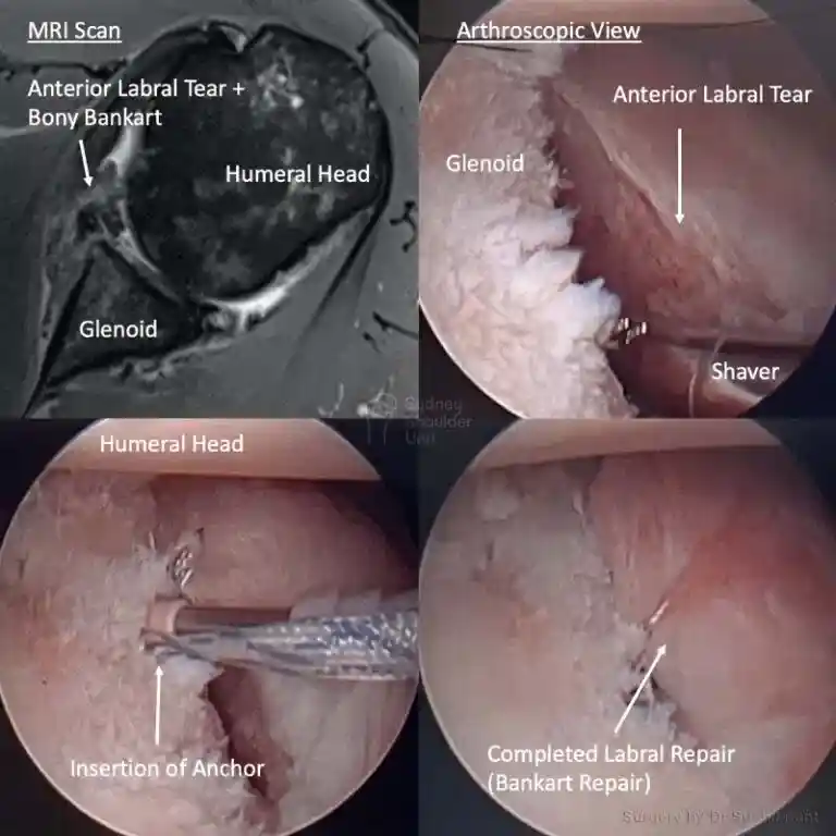
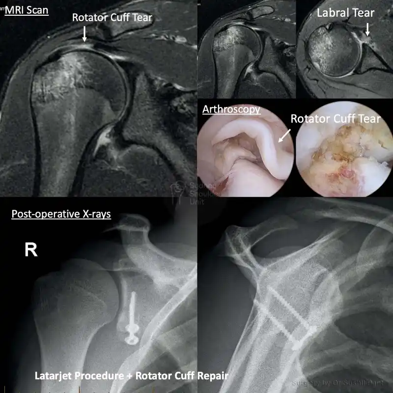
A MRI scan is almost always obtained for shoulder dislocations to look for labrum for tears  (anterior and posterior), SLAP tears, rotator cuff tears, cartilage surfaces, hill sachs and bony bankart lesions. In some cases it may be necessary to obtain a CT scan with 3D reconstructions to look at any bone loss on the glenoid or humeral head in more detail.

Medical diagram showing a human shoulder joint. The image highlights a Bankart lesion in the lower part of the labrum

Non-Surgical

Chronic (long-standing) shoulder instability is often first treated with non-surgical options. Acute shoulder dislocations in the majority of cases can also be treated non-surgically.  These may include physiotherapy, activity modification, and pain killers (NSAIDs).

Physiotherapy plays a key role in strengthening shoulder muscles and working on shoulder control (scapula control) can increase stability. You may need to continue with physiotherapy for some time to benefit. Immobilising your shoulder in a sling after dislocation should be minimised after the first few weeks; prolonged immobilisation in sling may lead to weakness of the shoulder muscles and worsen shoulder instability.

Surgical

Surgery is a good option for those who have failed to improve with time, physiotherapy, and activity modification.

In some cases surgery is a better option from the outset and these factors will be closely assessed by Dr Pant; some key considerations for early shoulder stabilisation surgery are:

  1. Age at first dislocation (<25)
  2. Overhead contact sport
  3. High-level athlete (contact sport +/- overhead)
  4. Glenoid (socket) bone loss
  5. Humeral head bone loss (Hill Sachs lesion)
  6. Rotator cuff tear with shoulder dislocation

There are two types of shoulder stabilisation surgeries:

  1. Arthroscopic (Keyhole) soft tissue repair; this includes things like a labral repair (bankart repair), SLAP repair, and remplissage procedure.
  2. Open bone block procedure (Latarjet); this is for those with significant bone loss (on the glenoid or humeral head) or high demand overhead athletes who take part in contact sport (rugby, AFL, soccer etc); during a latarjet procedure, the coracoid bone is transferred to the front on the socket via a split in the subscapularis muscle. It is a very successful procedure and provides the best. shoulder stabilisation possible.

The unstable shoulder

Repeat anterior instability/dislocations can lead to a fear of using the shoulder (not “trusting” it) and reduction in function. Patients may avoid positions of “apprehension”.

The painful shoulder

A labral tear at the back of the shoulder (Posterior)  may cause ongoing shoulder pain and difficulty sleeping on the affected side. Posterior labral tears usually do better with surgery. Tears of the Anterior Labrum usually lead to “apprehension” and loss of “trust” for fear of dislocation (more than pain per se)

Return to sport

Active individuals who wish to return to sport should consider surgery to repair torn Labrum. This will allow the ligaments to heal in the correct position. A torn Labrum, without surgery, will not heal itself.
The published return to sport rate after shoulder stabilisation surgery (key-hole) is 70-90%. With stabilisation surgery, patients are 5-6 times more likely to return to play than without surgery.

Anterior Tear

Patients will have apprehension with the hand in the upper outer quadrant
Inability to play overhead contact sports without instability +/- pain

Posterior Tear

Patients will have pain +/- apprehension with hand in the upper inner quadrant

Prevent post-traumatic arthritis

  • Repeat shoulder dislocations may damage the cartilage in the shoulder joint; this, in turn, leads to a higher risk of arthritis.
  • The published literature supports that shoulder stabilisation will reduce the risk of post-traumatic arthritis.
  • This is especially important in younger patients.
  • The procedure is under a General Anaesthetic where you will be positioned on your side and the affected arm secured in a special arm holder.
  • The shoulder is then prepared with a special antiseptic solution (dark pink in colour) and the surgical drapes are applied to maintain a sterile field at all times.
  • Small key-hole incisions (about 1cm) are made and the camera and surgical instruments are placed inside the shoulder. Usually 2-3 incisions are enough to carefully repair the Labrum
  • A thorough 15-point examination of the shoulder joint and torn Labrum is made via the arthroscopic camera and pictures are taken to document findings.
  • The torn Labrum is carefully prepared, scar tissue is cleaned up and stitches are placed around the Labrum which are in turn anchored back to the socket (2.9mm anchors). Usually 2-3 anchors in the front and/or  2-3 anchors in the back of the shoulder are needed.
  • Any other unexpected injuries/tears are noted and repaired via key-hole at the same time if needed.
  • The key-hole incisions are closed with sutures, dressings applied and a custom sling used to position the arm correctly (depending on your type of surgery).
  • Dr Pant will then organise close follow up at regular intervals and liaise with your Team Doctor/Physiotherapist to maximise your recovery.

General Shoulder Surgery Risks

Surgery is a carefully choreographed process and you are being treated by a sub-specialist shoulder surgeon and a highly experienced team; however, all surgeries inherently carry some risk of complications, such as (all are below 1%). The most common risks include:

  • Infection
  • Nerve injury
  • Bleeding

Specific Shoulder Surgery Risks

Frozen Shoulder

Frozen shoulder occurs when the shoulder joint capsule becomes inflamed, red and scarred – it initially presents with severe pain, then progresses to stiff ness and thaws out over many months

  • After routine shoulder surgery the risk of frozen shoulder is 5%. There is nothing you or your surgeon can do to reduce this risk
  • This risk is 10% if you have a history of Diabetes or Thyroid disorders.

Frozen shoulder progresses through three phases (which may each last 2-3 months):

PHASE 1 – Painful (even at rest and at night)

PHASE 2 – Stiff (reduction in movement)

PHASE 3 – Thawing out (increase in movement)

Natural history of frozen shoulder

Physiotherapy is not generally helpful in the first TWO PHASES of frozen shoulder. Once your shoulder starts to thaw out then you can work with your therapist within the arc of movement. Outcomes after a frozen shoulder:

  • Although it is a nuisance, a frozen shoulder will NOT change your outcome overall after surgery
  • In fact, in some cases, it is actually protective of your surgical repair
  • Occasionally patients need a second operation to “release” the frozen shoulder key-hole. This surgery is uncommon.

Reoperation and failure of shoulder surgery

When performing a shoulder reconstruction, your surgeon is using your own tissue to repair what is damaged. The weakest point in the repair is your own tissue. The longer the history of damage and frequency of dislocations/instability prior to surgery, the more likely the tissue quality is poor. There are three areas where your shoulder reconstruction may fail:

  1. Bone-Anchor interface. The anchors today are very high in quality and usually do not fail; sometimes if your bone is soft (older patients), the anchor may pull out of the bone.
  2. Anchor-Suture interface. This area is typically very strong and not a source of common failure.
  3. Suture-Tissue interface. This is the weakest point and the most common reason for failure of surgery (usually due to poor tissue quality)

There are special techniques in tissue management and suture choice to reduce failure rates.

There are three essential components to a good outcome from surgery:

  • Your surgeon
  • You as a patient
  • Your physical therapist

Dr Pant utilises the JPL pathway for most patients and this will form the basis of your rehabilitation after surgery; it will be modified to suit your individual circumstance.

The JPL pathway allows for self-directed, early passive shoulder range of movement. The full JPL pathway is available for download.

  • Passive – assisted with your other arm
  • Active – you move the affected arm independently

Sling

  • A sling is required for the first 6 weeks after your surgery
  • The sling is worn for a further 6 weeks outside of the house (to remind others you have had shoulder surgery.
  • The type of sling is selected specific to you and your shoulder surgery
  • Anterior labral repair – Normal sling
  • Posterior labral repair – ER sling (with a triangular wedge at the front)

When can I drive?

After shoulder surgery, you will be in a sling and you may be unable to drive for at least 6-8 weeks. This does vary depending on the complexity of your surgery and the patient. Some patients may be able to drive at 2-4 weeks, and some may not be able to drive for the full 12 weeks. This will be discussed at your follow up appointments.

Showering and getting dressed

  • You may take the sling off to shower
  • Lean forward and allow your arm to “dangle” to wash under your arms
  • The dressings applied are waterproof and you may shower with them on
  • Usually, before you are discharged from Hospital, your dressings are changed after your morning shower.
  • These “new” dressings are then kept intact until your review at 10-14 days
  • If the dressings start to peel at the edges – you may reinforce them
  • If the dressings discolour with discharge (yellow or green) you may have a wound infection – Do not be concerned just yet; please notify SSU (02 9215 6100 or admin@drpant.com.au) and we will give you a plan
  • When getting dressed, you may use your good arm to move your affected (operated arm) to place your arms through the sleeves carefully

Sleeping after Shoulder Surgery

  • Most patients find it difficult to lie flat after shoulder surgery
  • Consider using a few pillows to prop yourself up and sleep at a slight incline; this may be necessary for 4-6 weeks after surgery.
  • You should keep your sling on while asleep.

Elbow and hand movement

  • Keep your elbow and hands joints moving and supple for the duration of time you are in the sling.
  • The best time to move your elbow (into full extension and flexion) is when you are in the shower, just out of the shower and about to get dressed.
  • The published return to sport rate after shoulder stabilisation surgery (key-hole) is 70-90%. With stabilisation surgery, patients are 5-6 times more likely to return to play.
  • The risk of re-dislocation with overhead contact after soft tissue key-hole stabilisation is between 5-20% (depending on a number of factors).

Patient results

Shoulder Dislocation – Surfer

The gentleman is a keen surfer and dislocated his shoulder. He presented with instability symptoms and an X-ray that showed a bony bankart (fractured glenoid). He was unstable on examination and wished to return to his previous level of function overhead. 

The MRI scan confirmed an anterior labral tear with a bony bankart; in addition he had a posterior (back of the shoulder) labral tear and a SLAP tear. He had a corresponding Hill-Sachs lesion (fracture at the back of the Humeral head). 

He underwent an Arthroscopic bony bankart and labral repair as pictured below. We were able to repair the tissues back to their anatomical (original) position using suture anchors and achieved a nice “bumper” at the front of the shoulder. We also repaired the posterior labrum and the unstable SLAP lesion – all keyhole – a total of 3 small 1cm incisions around the shoulder. 

He commenced JPL therapy at two weeks and went on to strengthen his shoulder at 3 months. He is back surfing at six months post-surgery and happy with his outcome.

Shoulder Dislocation – Latarjet

After a shoulder dislocation some patients may be a candidate for a “bone transfer” procedure: a Latarjet. This is a good procedure for high level athletes, those who participate in overhead contact type sport, those with significant bone loss, and revision cases of failed previous “key-hole” stabilisation surgery.

This 16 year old high level wakeboarder dislocated his shoulder and sustained both a rotator cuff tear and an anterior labral tear. This is an uncommon injury pattern and presents unique decision making and surgical challenges.

Given his high level sporting ambitions and demands we repaired the Rotator Cuff and did a Latarjet procedure to treat his instability. He was in a sling for six weeks and commenced JPL rehabilitation at two weeks post surgery. He commenced formal Physiotherapy at 3 months, eventually regaining full ROM and function with his shoulder. He is back to his active lifestyle and riding waves around the waterways of Australia.

Shoulder Dislocation & Labral Tear

This 30 year old gentleman had a several year history of recurrent shoulder dislocations. He had exhausted a period of activity modification and physiotherapy. He was apprehensive to use his shoulder overhead, so he elected to proceed with surgery.

The images taken from surgery demonstrate a tear in the Labrum at the front of the shoulder. This has been carefully prepared and repaired back to the glenoid (socket) using 2.9mm anchors and suture-tape: achieving a nice “bumper” at the front of the shoulder joint.

He underwent JPL therapy and then physiotherapy to strengthen the shoulder. He no longer feels unstable and is back to participating in the sports he loves.

Request a call back

Enquiries between 8am-6pm (Mon-Fri) will be responded to within 30-60 minutes.

"*" indicates required fields

Alternatively, if you have any further questions or would like a consultation with Dr Pant get in touch:

Medicare Card Help

1. Medicare Number
2. Position on card
3. Expiry Date

Request a call back